Understanding Skin Cancer and Its Long-Term Effects
Skin cancer represents the most frequent form of cancer diagnosed worldwide. It develops when ultraviolet light from the sun damages the cellular DNA inside the skin, which causes cells to multiply out of control. Individuals with fair skin, light-colored eyes, numerous moles, or a family history possess a higher risk, though anyone can develop the condition. Early detection improves the clinical outlook and minimizes potential damage.
What Is Skin Cancer?
Physicians classify skin cancer into several categories, based on where abnormal cell growth begins. Basal cell carcinoma represents the most frequently diagnosed category. These growths typically appear on sun-exposed areas like the face, neck, and head. They can manifest as scar-like flat lesions, raised red patches, small pearly bumps, or open sores that fail to heal. The growths are fragile and may bleed after minor friction.
Squamous cell carcinoma originates in the flat cells comprising the skin’s top layer, and presents as reddish, scaly, or crusted patches on the face, ears, neck, and hands. Melanoma is a less common but more aggressive form of the disease. It develops in melanocytes, which are the cells responsible for skin pigmentation. It resembles a new spot or an existing mole that has changed in size, shape, or color.
What Are the Effects?
The long-term effects of skin cancer depend on the specific type, the location of the growth, and the stage at which a physician makes a diagnosis. Basal cell and squamous cell carcinomas typically grow at a slow pace. If left untreated, the abnormal cells can penetrate deeper layers of skin, underlying tissue, and bone. This penetration leads to physical disfigurement, localized tissue destruction, and scarring following eventual surgical removal. Melanoma carries a high risk of metastasis, which means cancer cells can spread from the initial site to other parts of the body. Patients who have had one skin cancer diagnosis possess a statistically higher likelihood of developing another growth.
How Is Cancer Treated?
Medical professionals determine the appropriate therapy based on the cancer’s form, stage, and bodily location. Dermatologists typically begin with a biopsy and examine a small tissue sample under a microscope. This confirms the presence and exact nature of the cancerous cells.
Mohs surgery serves as a highly effective outpatient procedure for localized carcinomas. A specialized surgeon removes the cancerous tissue layer by layer and examines each section microscopically until no abnormal cells remain. This precise method yields high success rates while preserving the maximum amount of healthy surrounding skin.
Other standard therapies exist for varying stages and types of the disease. Physicians might freeze the tumor, removing it after the tissue is destroyed. Alternative approaches include curettage and desiccation, which involve scraping the tissue and applying targeted heat.
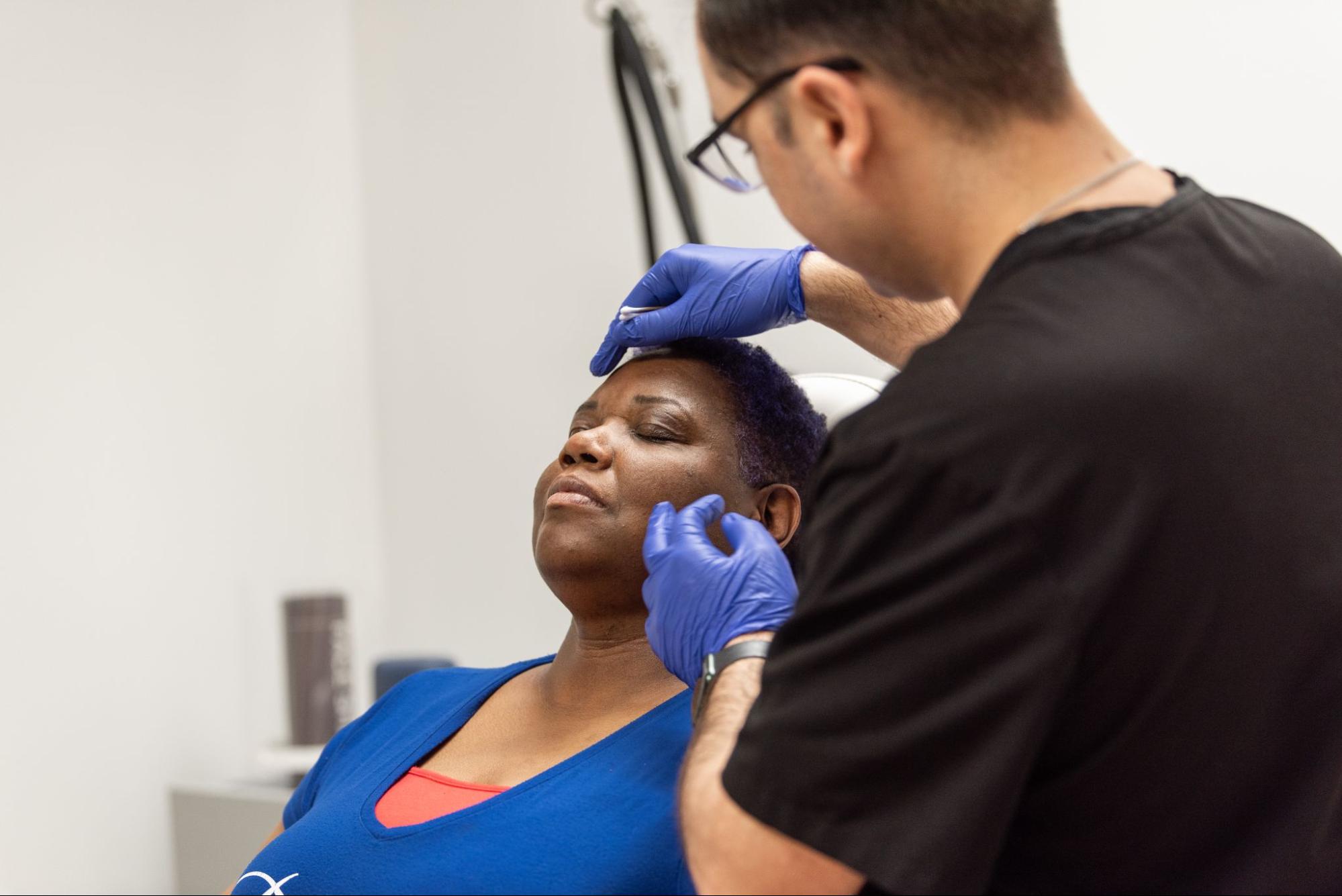
Consult With a Dermatologist
Catching cellular abnormalities in their earliest stages provides the highest probability of a favorable clinical outcome. Anyone noticing a changing mole, a new patch of discolored skin, or an unhealing sore should schedule a clinical evaluation. A dermatologist possesses the specialized training required to assess suspicious spots and design comprehensive treatment plans. Seeking professional medical guidance immediately upon noticing a visible dermatological change serves as an effective step in managing long-term health, helping to minimize disease progression.
- How to Sell Your House Fast in a Competitive Market
- How to Prepare Your Home for a Successful Sale: The Essential Steps
- Understanding Cash Home Buying in Detroit, Michigan: A Comprehensive Guide
- Exploring Homeownership in Columbus, GA: A Comprehensive Guide
- The Benefits of Selling Your House Quickly During Sudden Life Changes